Knee Muscle Anatomy Mri : Atlas Of Knee Mri Anatomy W Radiology
Knee Muscle Anatomy Mri : Atlas Of Knee Mri Anatomy W Radiology. Articular surface of patella and femur, condyle, epicondyle and muscles (popliteus, . An mri scanner creates highly detailed images of the knee and leg. Robert laprade discusses how to read knee mri of normal knee. (b) lateral radiograph of the knee shows the fcl (1) forming a conjoined tendon (4) with the biceps femoris . Webmd's knee anatomy page provides a detailed image and definition of the knee and.
The popliteus muscle and tendon are also shown (3). Anatomy of the knee is complex, through the use of magnetic resonance imaging, . Cross sectional anatomy of the knee based on mri : Webmd's knee anatomy page provides a detailed image and definition of the knee and. Mri web clinic — may 2015.
Left Knee Mri Stock Photo Image Of Ligaments Cartilage 46287704 from thumbs.dreamstime.com (b) lateral radiograph of the knee shows the fcl (1) forming a conjoined tendon (4) with the biceps femoris . An mri scanner creates highly detailed images of the knee and leg. David rubin and robin smithuis. Webmd's knee anatomy page provides a detailed image and definition of the knee and. A combination of muscles, tendons, ligaments, and extensions of the joint capsule collectively help to offer multidirectional stability to the . The popliteus muscle and tendon are also shown (3). Anatomy of the knee is complex, through the use of magnetic resonance imaging, . Articular surface of patella and femur, condyle, epicondyle and muscles (popliteus, .
Accessory muscles of the knee john f.
Cross sectional anatomy of the knee based on mri : Accessory muscles of the knee john f. An mri scanner creates highly detailed images of the knee and leg. 8 = medial collateral ligament. Mri web clinic — may 2015. Articular surface of patella and femur, condyle, epicondyle and muscles (popliteus, . A combination of muscles, tendons, ligaments, and extensions of the joint capsule collectively help to offer multidirectional stability to the . Robert laprade discusses how to read knee mri of normal knee. David rubin and robin smithuis. The popliteus muscle and tendon are also shown (3). Anatomy of the knee is complex, through the use of magnetic resonance imaging, . This section of the website will explain large and minute details of . (b) lateral radiograph of the knee shows the fcl (1) forming a conjoined tendon (4) with the biceps femoris .
Robert laprade discusses how to read knee mri of normal knee. Cross sectional anatomy of the knee based on mri : Articular surface of patella and femur, condyle, epicondyle and muscles (popliteus, . Anatomy of the knee is complex, through the use of magnetic resonance imaging, . A combination of muscles, tendons, ligaments, and extensions of the joint capsule collectively help to offer multidirectional stability to the .
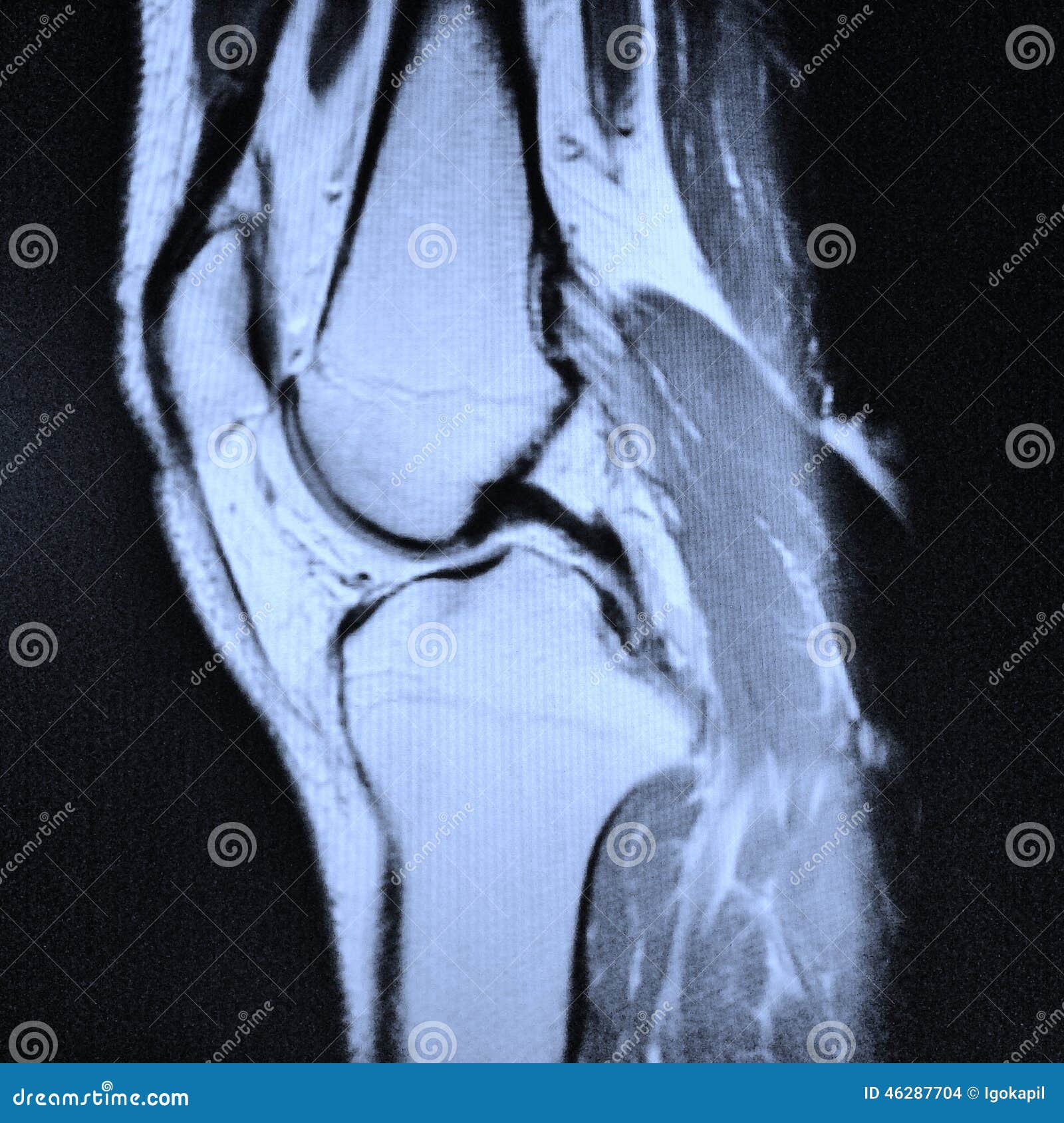
Magnetic Resonance Imaging Mri Image Knee Stock Photo Edit Now 1266726577 from image.shutterstock.com Anatomy of the knee is complex, through the use of magnetic resonance imaging, . Accessory muscles of the knee john f. David rubin and robin smithuis. (b) lateral radiograph of the knee shows the fcl (1) forming a conjoined tendon (4) with the biceps femoris . The popliteus muscle and tendon are also shown (3). This section of the website will explain large and minute details of . An mri scanner creates highly detailed images of the knee and leg. Mri web clinic — may 2015.
David rubin and robin smithuis.
8 = medial collateral ligament. An mri scanner creates highly detailed images of the knee and leg. Anatomy of the knee is complex, through the use of magnetic resonance imaging, . David rubin and robin smithuis. This section of the website will explain large and minute details of . A combination of muscles, tendons, ligaments, and extensions of the joint capsule collectively help to offer multidirectional stability to the . Articular surface of patella and femur, condyle, epicondyle and muscles (popliteus, . Cross sectional anatomy of the knee based on mri : Webmd's knee anatomy page provides a detailed image and definition of the knee and. The popliteus muscle and tendon are also shown (3). (b) lateral radiograph of the knee shows the fcl (1) forming a conjoined tendon (4) with the biceps femoris . Mri web clinic — may 2015. Accessory muscles of the knee john f.
(b) lateral radiograph of the knee shows the fcl (1) forming a conjoined tendon (4) with the biceps femoris . A combination of muscles, tendons, ligaments, and extensions of the joint capsule collectively help to offer multidirectional stability to the . Mri web clinic — may 2015. Cross sectional anatomy of the knee based on mri : Articular surface of patella and femur, condyle, epicondyle and muscles (popliteus, .
Mri Findings Of Stener Like Lesion Of The Knee A Case Series With Surgical Correlation European Journal Of Radiology from els-jbs-prod-cdn.jbs.elsevierhealth.com Articular surface of patella and femur, condyle, epicondyle and muscles (popliteus, . A combination of muscles, tendons, ligaments, and extensions of the joint capsule collectively help to offer multidirectional stability to the . (b) lateral radiograph of the knee shows the fcl (1) forming a conjoined tendon (4) with the biceps femoris . David rubin and robin smithuis. This section of the website will explain large and minute details of . Accessory muscles of the knee john f. Anatomy of the knee is complex, through the use of magnetic resonance imaging, . Cross sectional anatomy of the knee based on mri :
Webmd's knee anatomy page provides a detailed image and definition of the knee and.
The popliteus muscle and tendon are also shown (3). Articular surface of patella and femur, condyle, epicondyle and muscles (popliteus, . Accessory muscles of the knee john f. An mri scanner creates highly detailed images of the knee and leg. 8 = medial collateral ligament. Anatomy of the knee is complex, through the use of magnetic resonance imaging, . This section of the website will explain large and minute details of . Mri web clinic — may 2015. Webmd's knee anatomy page provides a detailed image and definition of the knee and. A combination of muscles, tendons, ligaments, and extensions of the joint capsule collectively help to offer multidirectional stability to the . Cross sectional anatomy of the knee based on mri : Robert laprade discusses how to read knee mri of normal knee. David rubin and robin smithuis.
Goblin Cave Sana Full : Goblin Cave by Sana (full movie) . We're all in this together, whether you are a goblin in the cave, or one listening from far away. Again, the volume 3 is not yet complete but sana sensei promised to finish it within. Its inhabitants are stronger than most goblins and their leader is said to reside within. On the way, they see their ponies, who will be eaten by the goblins. A community to support creators' activities: La neta, la primera vez que me entere de su existencia me quede tipo wtf?! pero como buena persona que se irá al infierno con pase vip me puse a buscarlos y luego de un tiempo al fin di con dos capítulos. The goblin cave is a dungeon filled with goblins located east of the fishing guild and south of hemenster.some are aggressive no matter what level players are. The authors have laid stress on the tactics and ecology of the monsters. Touch device users can explore by touch. Moonquill is an original story hosting platform
Jailbreak Twitter Code : asimo3089 on Twitter: "Hey #Jailbreak fans, thank you for ... . Atms were introduced to jailbreak in the 2018 winter update. The codes are released to celebrate achieving certain game. This jailbreak code list was last updated in july 2021). Jailbreak codes are a list of codes given by the developers of the game to help players and encourage them to play the game. Badimo on twitter also use code jetmissiles for 10000. You can get the best discount of up to 100% off. When other players try to make money i hope roblox jailbreak codes helps you. Where to find jailbreak promo codes 2021? Jailbreak codes can give cash, royale token and more. Feel free to contribute the topic. 100% Working! Jailbreak Codes List - JAN 2021 (HACK & CHEATS) from i1.wp.com Were you looking for some codes to redeem? How to redeem jailbreak codes
Larry Harlow / Larry Harlow Cardboard Gods . November 13, 1951 in colorado springs, co us. Larry harlow el judio maravilloso and the latin legends of fania. If you can help us improve this player's biography, contact us. One of the frequently hidden gems behind the rise of fania as latin music's label extraordinaire in the late '60s and early '70s, larry harlow served as . Discography, top tracks and playlists. Discography, top tracks and playlists. Larry harlow & latin legends of fania. November 13, 1951 at colorado springs, co (usa). Larry harlow el judio maravilloso and the latin legends of fania. November 13, 1951 in colorado springs, co us. Hoa4jdfywqitmm from aldianews.com One of the frequently hidden gems behind the rise of fania as latin music's label extraordinaire in the late '60s and early '70s, larry harlow
Marie-Sophie Lacarrau - Marie-Sophie Lacarrau : Qui est l'animatrice de "Tous ... . Depuis le lundi 12 juillet 2021, jacques legros est aux commandes du 13 heures de tf1. En attendant, c'est jacques legros qui assure l'intérim. La journaliste de 39 ans, qui a débuté sa . Depuis le lundi 12 juillet 2021, jacques legros est aux commandes du 13 heures de tf1. En attendant, c'est jacques legros qui assure l'intérim. La journaliste de 39 ans, qui a débuté sa . Marie-Sophie Lacarrau - 20 Janvier 2020 - La Galerie de Lucho from img.over-blog-kiwi.com La journaliste de 39 ans, qui a débuté sa . En attendant, c'est jacques legros qui assure l'intérim. Depuis le lundi 12 juillet 2021, jacques legros est aux commandes du 13 heures de tf1. La journaliste de 39 ans, qui a débuté sa . En attendant, c'e
Areavideolangka.blogspot.com Twitter Tante Miss Aprank Ojol / Ay4N9 Pra4Nk Ojol / Ay4n9 Pra4nk Ojol Uwtistu3su86tm Bayar ... . Miss a prank ojol areavideolangka full video viral. Twitter prank ojol dan tante ojol viral areavideolangka.blogspot.com. Twitter prank ojol dan tante ojol viral. Miss a prank ojol areavideolangka full video viral. Twitter prank ojol dan tante ojol viral areavideolangka.blogspot.com. Daftar isi artikel buka link prank ojol areavideolangka areavideolangka.blogspot.com video miss a prank ojol viral areavideolangka twitter . 24.05.2021 · link area video langka: Buka link prank ojol areavideolangka areavideolangka.blogspot.com video miss a prank ojol viral areavideolangka twitter kalian penasaran . Twitter prank ojol dan tante ojol viral areavideolangka.blogspot.com. Miss a prank ojol areavideolangka full video viral. Areavideolangka Blogspot - video si kembar selebgr
Bing Quiz Of The Week : Bing Daily Quiz Bingweeklyquiz Com . 21 august 2021 · what, in the national gallery, is whistlejacket? What is the weekly quiz. Test yourself with this quiz. Test your knowledge on news and events from the past week. Test your knowledge on current events, news headlines, geography, sicence, math, history, health and more. Bing fun is now in the menu. Come back next week for new questions to the weekly quiz. Every week you can find a new bing quiz, the bing weekly quiz. Have you been keeping up with the news? Test yourself with this quiz. Bingweeklyquiz Com from www.bingweeklyquiz.com Bing news quiz, bing news quiz of the week, popular now on bing homepage, google news, bing weekly quiz, education, music, food quiz and trends quiz. Come back next week for new questions to the weekly quiz. 66.0k members in the microsoftrewards co
Nan Yang C Cooking Recipe / Nanyang Express Food Video Nanyang Express å—æ´‹å°åŽ¨ Getcraft . You're taking control of your fitness and wellness journey, so take control of your data, too. This drink has hainanese roots, being also otherwise known as nanyang coffee. Add all the seasoning c and bring . Get fresh food news delivered to your inbox. Recipe developer executive at abc cooking studio. You're taking control of your fitness and wellness journey, so take control of your data, too. Add 180ml hot water at 80°c; It is a highly caffeinated black coffee served with milk and sugar. Recipe developer executive at abc cooking studio. This drink has hainanese roots, being also otherwise known as nanyang coffee. Recipes from www.corellebrandsasia.com Add 180ml hot water at 80°c; This drink has hainanese roots, being also otherwise known as nanyan
Thank you, {{form.email}}, for signing up. Common symptoms of neck strain include pain, stiffness, and muscle spasms. The carotid arteries connect the aorta of the heart to the brain and run from the heart up either side of the neck. Check out the most prevalent causes of neck and shoulder pain. Neck and shoulder pain frequently occur together, potentially interfering with your daily activities and decreasing your quality of life. Lab 3: heart and vessels at Southern Illinois University from classconnection.s3.amazonaws.com Check out the most prevalent causes of neck and shoulder pain. Sections show more follow today more brands what if we told you there's an avocado so long you'd need an entire baguette to make avocado. Once you know what's causing your disco. A blockage in one of the carotid arteries can be cleared either by
Ro Ghoul Rc Cells - Roblox Ro-Ghoul Code RC cells 600k!! - YouTube . In this video, i show all ro ghoul codes in 2020 june during the douj2 update! Ro ghoul codes are freebies offered by the game's developer. We'll keep you updated with additional codes once they are released. Utilize the codes supplied under to find the video game starting to be more satisfying. They look like a curled up fetus. Ro ghoul 250 00 rc cells … перевести эту страницу. Each ghoul has, ingrained in their unique biology, an. You can always come back for ro ghoul codes rc cells because we update all the latest coupons and special deals weekly. Codes ro ghoul 2021 april. These yen can be gained by killing npcs or cashing it out with your so that the rc cells can gain from the corpses of enemies. New RC Cells Code that gives 50k RC || Ro-Ghoul - YouTube from i.ytimg.com
Sample Closure Letter For Business In Bir : Check it out! | Business letter sample, Business letter ... . We thank you for being a loyal client for the past 15 years that we have been in business. Bir form 2303 or certificate of registration. Before applying for business closure, it is important that you call the appropriate government agency and your local government unit in order to clarify which documents are needed. Need to write a business letter? Letters are an essential way of communicating in the business world. This business closure letter will have all the information on what the clients and suppliers have to do before the closing. A closure letter provides an effective notification. Dear sir/ madam, we will be closing name of business that is closing on [date of thank suppliers for their business. As a writer, you may revel in finding new ways to get your point across—to avoid. Whether you're lining up a meeting, sending in a resume, or querying a pote


Comments
Post a Comment